



Loading page…
CT Scan and Radiology
Price Starts From | No Hidden Charges
Loading price…
₹3,500/*
*Prices may vary by city and chosen centre. Inclusive of radiologist report. Contrast scans priced separately. No hidden charges.
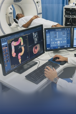
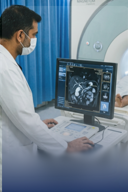
A Brain CT Scan (Computed Tomography of the Brain) is a rapid, cross-sectional imaging investigation that produces detailed images of the brain, skull, and intracranial structures using X-rays. Unlike a standard X-ray, CT generates thin-slice images from multiple angles — reconstructed into a complete three-dimensional picture of the brain. A Brain CT is the fastest and most widely available imaging tool for brain emergencies — including stroke, intracranial haemorrhage, and head trauma. It can be completed in under 5 minutes for the scan itself, making it essential in time-critical situations. Two types of Brain CT: * Plain (NCCT) Brain CT: No contrast injection required — the standard scan for most emergencies, including stroke, bleeding, and head injury. * Contrast (CECT) Brain CT: A contrast dye is injected intravenously before scanning — used for tumour detection, infection (abscess), and blood vessel abnormalities. Requires 4 hours fasting and a kidney function check (creatinine) before contrast administration. CT vs MRI: CT is faster and better for acute bleeding and bone injury. MRI provides better soft tissue and white matter detail — used for stroke characterisation, tumour staging, and when CT findings are inconclusive.
Key benefits of this package include:
Ischaemic stroke — loss of normal brain density in a vascular territory
Haemorrhagic stroke — intracerebral haemorrhage (hyperdense blood)
Subarachnoid haemorrhage (SAH) — blood in the spaces around the brain (thunderclap headache)
Subdural haematoma — blood between brain and dura, typically from falls
Extradural haematoma — blood between skull and dura, from arterial tear after head trauma
Cerebral contusion — brain bruising from trauma
Skull fracture — including depressed fractures and base of skull fractures
Primary brain tumours — glioma, meningioma, pituitary adenoma
Brain metastases — secondary tumours from lung, breast, or other primary cancers
Brain abscess — ring-enhancing lesion on contrast CT
Lymphoma — in immunocompromised patients
Cerebral oedema — brain swelling from any cause
Hydrocephalus — enlarged ventricles from CSF obstruction or overproduction
Cerebral atrophy — generalised brain volume loss (dementia workup)
Calcifications — physiological or pathological calcium deposits
Midline shift — displacement of brain structures from a mass or oedema
Sinusitis — incidental finding of air-sinus disease

Explore other scans and imaging services — book online or request a call back.
Pathology tests that pair well with your imaging — book home collection online.